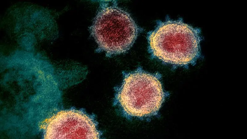
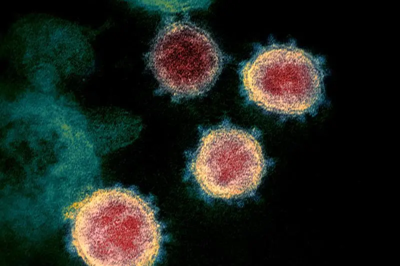
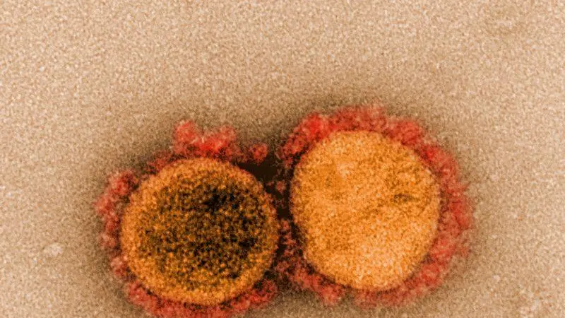
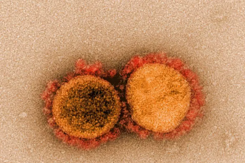
An isolate from the first U.S. case of COVID-19, formerly known as 2019-nCoV or novel coronavirus, is seen in a transmission electron microscopic image obtained from the Centers for Disease Control (CDC) in Atlanta, Georgia, U.S. March 10, 2020. CDC/Hannah A Bullock and Azaibi Tamin/Handout via REUTERS.
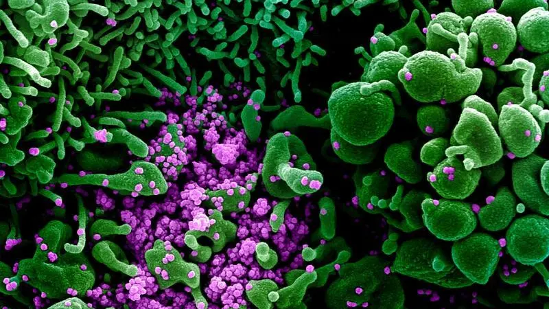
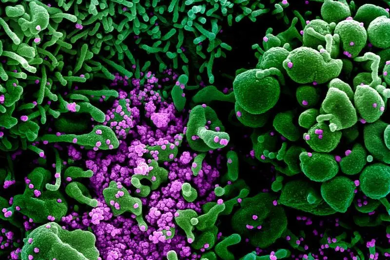
An undated colorized scanning electron micrograph of an apoptotic cell (green) heavily infected with SARS-COV-2 virus particles (purple), also known as novel coronavirus, the virus which causes COVID-19, isolated from a patient sample. NIAID Integrated Research Facility (IRF)/Handout via REUTERS.
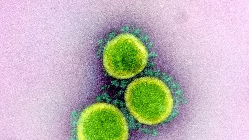
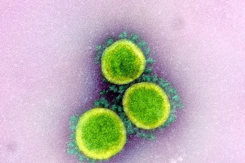
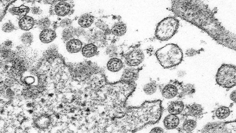
An undated transmission electron micrograph of SARS-CoV-2 virus particles, also known as novel coronavirus, the virus that causes COVID-19, isolated from a patient. NIAID Integrated Research Facility (IRF)/Handout via REUTERS.
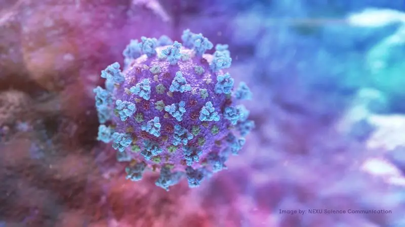
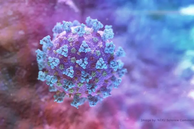
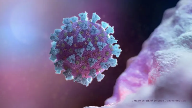
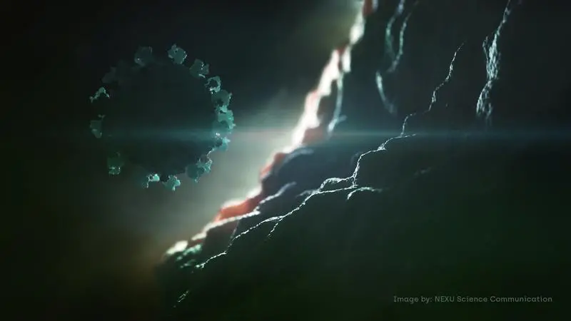
A computer image created by Nexu Science Communication together with Trinity College in Dublin, shows a model structurally representative of a betacoronavirus which is the type of virus linked to COVID-19, better known as the coronavirus linked to the Wuhan outbreak, shared with Reuters on February 18, 2020. NEXU Science Communication/via REUTERS
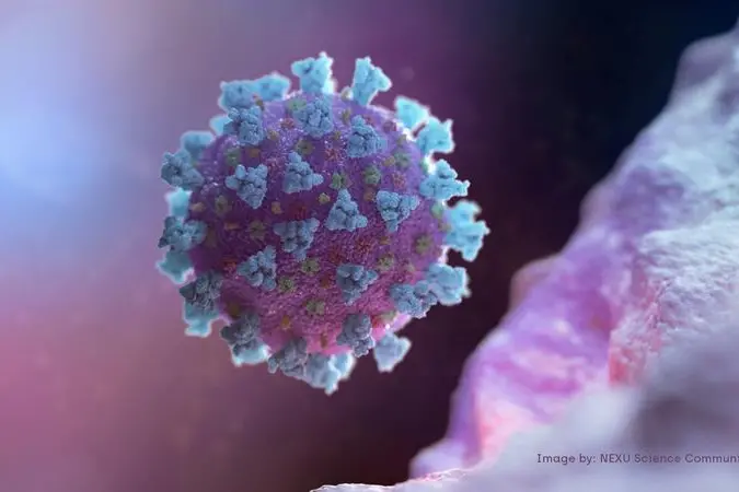
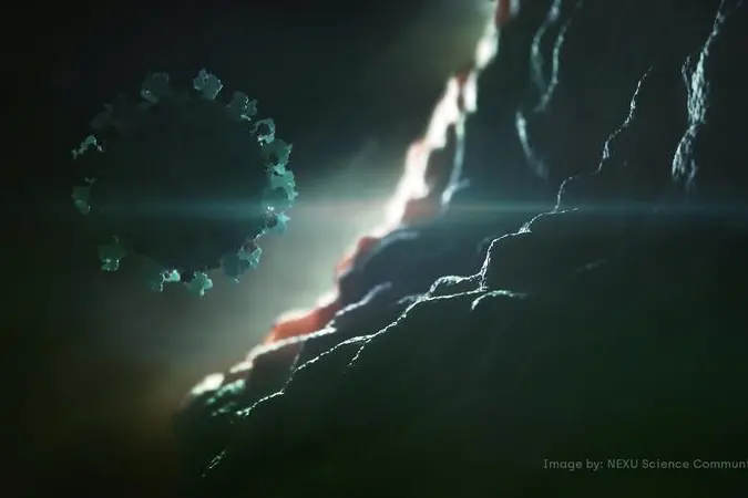
A computer image created by Nexu Science Communication together with Trinity College in Dublin, shows a model structurally representative of a betacoronavirus which is the type of virus linked to COVID-19, better known as the coronavirus linked to the Wuhan outbreak, shared with Reuters on February 18, 2020. NEXU Science Communication/via REUTERS
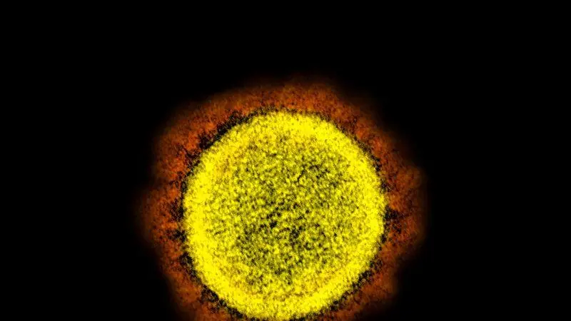
An undated scanning electron microscope image shows SARS-CoV-2 (yellow), also known as novel coronavirus, the virus that causes COVID-19, isolated from a patient in the U.S., emerging from the surface of cells (blue/pink) cultured in the lab. NIAID-RML/Handout via REUTERS.
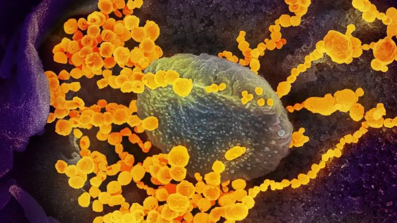
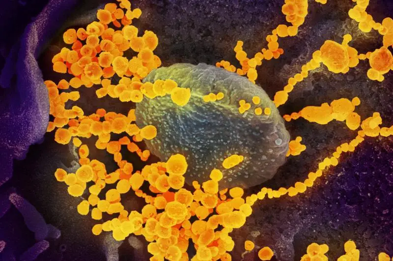
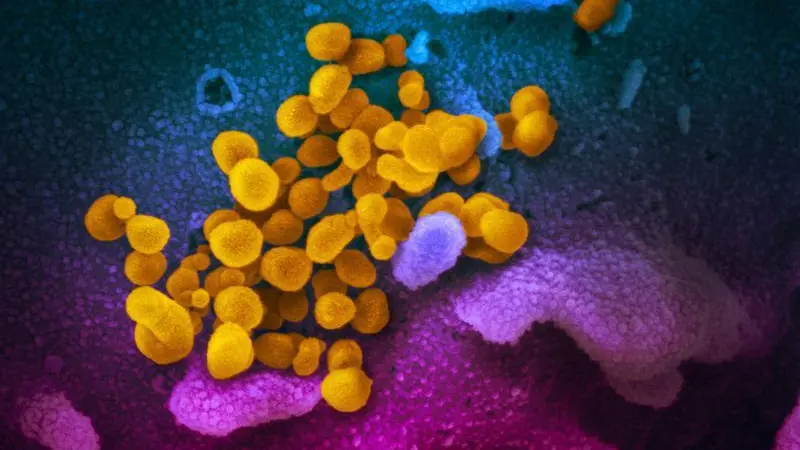
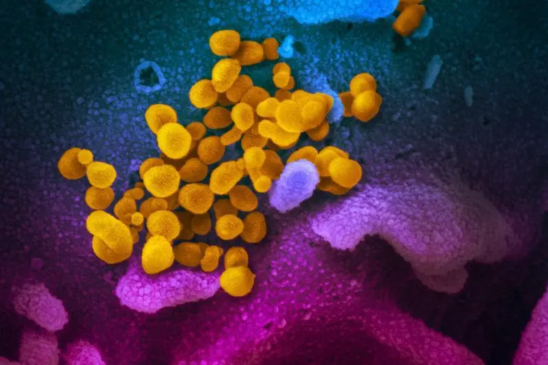
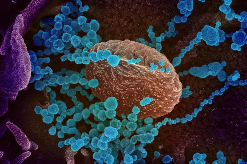
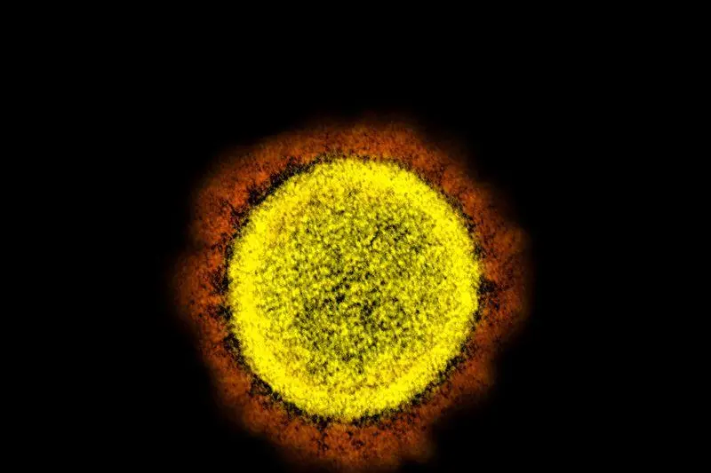
An undated scanning electron microscope image shows SARS-CoV-2 (round gold objects), also known as novel coronavirus, the virus that causes COVID-19, emerging from the surface of cells cultured in the lab and isolated from a patient in the U.S. NIAID-RML/Handout via REUTERS.
This undated transmission electron microscope image shows SARS-CoV-2, also known as novel coronavirus, the virus that causes COVID-19, isolated from a patient in the U.S. Virus particles are shown emerging from the surface of cells cultured in the lab. The spikes on the outer edge of the virus particles give coronaviruses their name, crown-like. NIAID-RML/Handout via REUTERS.
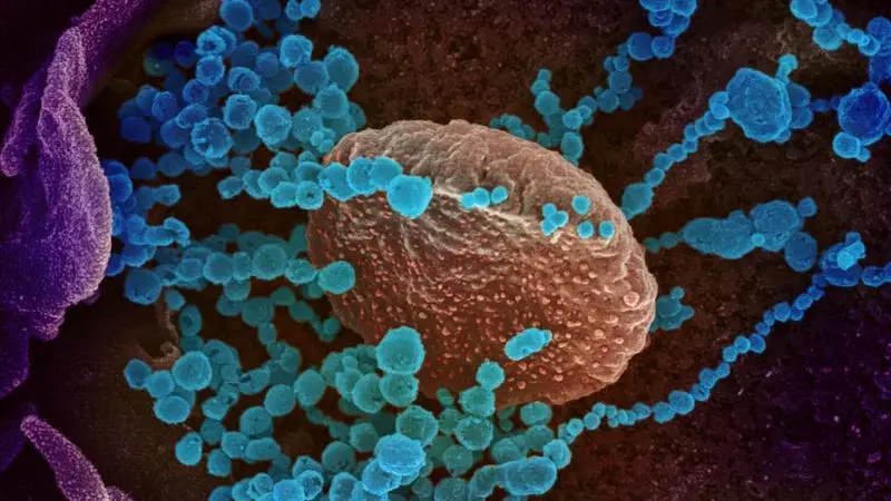
This scanning electron microscope image shows SARS-CoV-2 (round blue objects), also known as novel coronavirus, the virus that causes COVID-19, emerging from the surface of cells cultured in the lab which was isolated from a patient in the U.S. NIAID-RML/Handout via REUTERS.
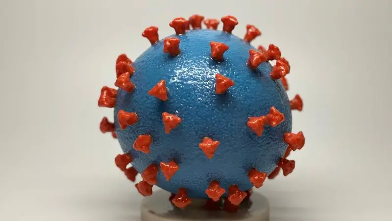
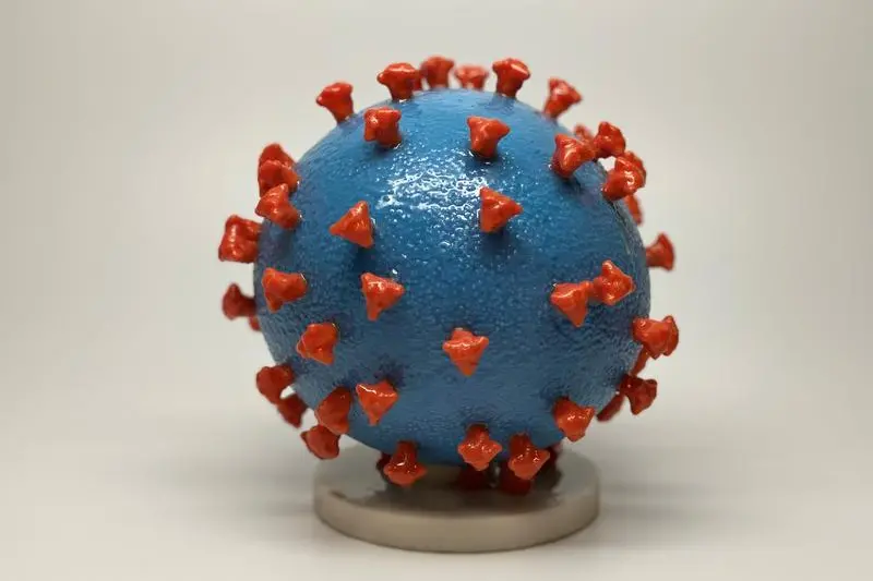
An undated photo shows a 3-D print of a SARS-CoV-2 particle, also known as novel coronavirus, the virus that causes COVID-19. The virus surface (blue) is covered with spike proteins (red) that enable the virus to enter and infect human cells. The spikes on the surface of coronaviruses give this virus family its name ? corona, which is Latin for ?crown,? and most any coronavirus will have a crown-like appearance. NIH/Handout via REUTERS.
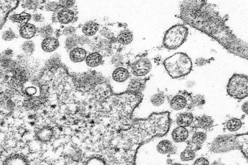
An undated transmission electron micrograph of a SARS-CoV-2 virus particle, also known as novel coronavirus, the virus which causes COVID-19, isolated from a patient. NIAID Integrated Research Facility (IRF)/Handout via REUTERS.
A computer image created by Nexu Science Communication together with Trinity College in Dublin, shows a model structurally representative of a betacoronavirus which is the type of virus linked to COVID-19, better known as the coronavirus linked to the Wuhan outbreak, shared with Reuters on February 18, 2020. NEXU Science Communication/via REUTERS
An undated transmission electron micrograph of SARS-CoV-2 virus particles, also known as novel coronavirus, the virus that causes COVID-19, isolated from a patient. NIAID Integrated Research Facility (IRF)/Handout via REUTERS.
Up close with the coronavirus